The web site presents a collection of normal and pathological digitalized histological sections that can be easily navigated and observed at various magnifications
...continue to readThe Cell: An Image Library™ is a freely accessible, easy-to-search, public repository of reviewed and annotated images, videos, and animations of cells from a variety of organisms, showcasing cell architecture, intracellular functionalities, and both normal and abnormal processes. The purpose of this database is to advance research, education, and training, with the ultimate goal of improving human health.
...continue to readThe Atlas of histological images is part of the Anatomy Atlases, a digital library of anatomy information curated by Ronald A Bergman.
...continue to readPathorama provides you with high quality images and virtual slides for teaching and self-instruction covering a wide range of topics in all subspecialities of surgical pathology and cytology. Various courses, slide seminars, quizzes, and learning games for both students, surgical pathologists, and cytopathologists are available.
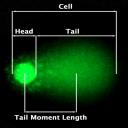
...continue to readThis site is dedicated to explain concepts and technology behind the Comet Assay to detect DNA damage.
...continue to readConfocal Microscopy tutorial (University of Helsinki) does not copy all the theoretical and technical details of confocal microscopy, instead, it delineate the outline about how confocal microscope works, and some basic concepts that are important for a user to understand or decide which button to press, which knob to turn, etc..
...continue to read
Add to my favorites
Remove from my favorites
protocols for 2D and 3D quantification in microscopic images
Category: Imaging software, Imaging tutorials
Subcategories: Image analysis
Microscopy-based imaging is booming and the need for tools to retrieve quantitative data from images is urgent. This book provides simple but reliable tools to generate valid quantitative gene expression data, at the mRNA, protein and activity level, from microscopic images in relation to structures in cells, tissues and organs in 2D and 3D. Volumes, areas, lengths and numbers of cells and tissues can be calculated and related to these gene expression data while preserving the 2D and 3D morpholo
...continue to readIt allows to examine pathological sections at different magnifications as under a microscope
...continue to readPage: 1


